Our Summary
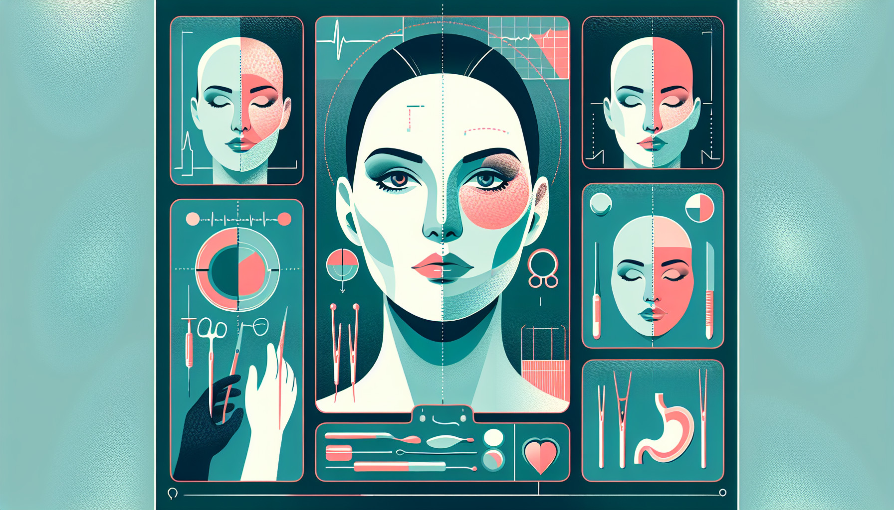
This research paper talks about a new method to improve the treatment of a craniofacial bone disorder called fibrous dysplasia, which affects the upper jaw and cheekbone. The traditional treatment involves shaving off some bone, but it’s hard to know exactly how much to remove.
The authors have come up with a new technique using 3D printing and screws. They first use a CT scan to get an image of the bone, then they use 3D printing technology to create a guide. They put screws of specific lengths into the bone during surgery, which helps them know exactly how much bone to shave off.
This method makes the surgery quicker and easier, and also reduces the patient’s exposure to radiation from repeated CT scans. It’s also cheaper. The results have been good, with patients looking better after surgery than with the traditional method.
FAQs
- What is the optimal alternative treatment for craniofacial fibrous dysplasia affecting the maxilla and zygoma?
- How does the new method using three-dimensional printing technology improve the bone-shaving procedure?
- What are the benefits of this new method of bone shaving compared to current practices?
Doctor’s Tip
A helpful tip a doctor might give a patient undergoing craniofacial surgery is to follow all pre-operative instructions carefully, including any specific dietary restrictions or medication guidelines. It is also important to discuss any concerns or questions with the surgical team beforehand to ensure a successful outcome. Additionally, post-operative care, such as proper wound care and follow-up appointments, should be followed diligently to promote healing and recovery.
Suitable For
Patients who are recommended for craniofacial surgery typically include those with craniofacial fibrous dysplasia affecting the maxilla and zygoma. This condition may cause facial deformities, asymmetry, and functional issues. In some cases, traditional bone-shaving procedures may not be effective or may result in unpredictable outcomes. In such cases, the novel method described above using three-dimensional printing technology and screws as a guide may be recommended for more precise and effective results. This method may be particularly beneficial for patients who require accurate removal of bone in specific areas of the face to improve both function and aesthetics.
Timeline
Before craniofacial surgery:
- Patient is diagnosed with craniofacial fibrous dysplasia affecting the maxilla and zygoma.
- Consultation with a craniofacial surgeon to discuss treatment options.
- Preoperative imaging studies such as CT scans are done to assess the extent of the condition.
After craniofacial surgery:
- Three-dimensional printing technology is used on CT scans to plan the bone-shaving procedure.
- Screws of predetermined lengths are implanted into surgical sites as a guide for the bone shaving.
- Surgeon performs the bone shaving within the visual field using the screws as a reference.
- Operation time is shortened and less effort intensive, with reduced risk of radiation exposure and low cost.
- Postoperative follow-up appointments to monitor healing and cosmetic outcomes of the surgery.
What to Ask Your Doctor
- What specific areas of my craniofacial structure will be targeted with the bone-shaving procedure?
- How will three-dimensional printing technology be utilized in my surgery?
- What is the expected outcome in terms of cosmetic improvements?
- What are the potential risks and complications associated with this procedure?
- How long is the recovery process expected to be?
- Will I require any additional treatments or follow-up appointments after the surgery?
- How experienced are you in performing craniofacial surgery using this novel method?
- Are there any alternative treatment options available for my condition?
- Are there any specific pre-operative or post-operative instructions I should follow?
- Can you provide me with any before and after photos of previous patients who have undergone similar craniofacial surgeries using this method?
Reference
Authors: Kang SJ, Oh MJ, Jeon SP. Journal: J Craniofac Surg. 2015 Sep;26(6):1977-8. doi: 10.1097/SCS.0000000000001953. PMID: 26192030