Our Summary
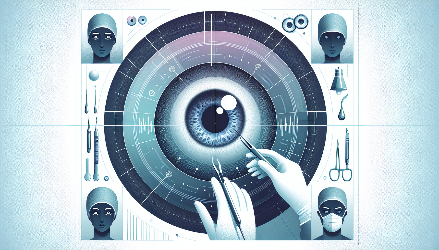
This study aimed to assess the effectiveness of a particular type of retinal imaging (Optomap 200Tx) for identifying potential issues in the retina before a patient undergoes refractive eye surgery. The researchers studied 208 eyes from 109 people who were about to have this surgery. They used the Optomap along with two other methods of eye examination.
They found that just over a third of the eyes (73 out of 208) had some kind of peripheral retinal issue when examined by a specialist. The Optomap was able to identify these issues in 78.1% of cases, with a specificity rate (i.e., its ability to correctly identify those without the condition) of 60%. Accuracy varied between different people interpreting the results, ranging from 72% to 87%, with the highest accuracy seen in those with previous retinal training.
In simple terms, the Optomap was quite good at spotting potential problems in the retina before surgery, particularly when the results were interpreted by someone with appropriate training. It’s a convenient and quick method for checking the health of the retina in patients who are short-sighted.
FAQs
- What is the purpose of using Optomap 200Tx in retinal imaging?
- How effective is the Optomap 200Tx in identifying peripheral retinal issues before refractive eye surgery?
- Does the accuracy of the Optomap 200Tx vary based on who is interpreting the results?
Doctor’s Tip
A doctor might advise a patient undergoing retinal surgery to consider getting a retinal imaging scan, such as an Optomap, before the procedure. This can help identify any potential issues in the retina that may affect the success of the surgery. Additionally, the doctor may recommend regular follow-up appointments and monitoring of the retina post-surgery to ensure optimal outcomes.
Suitable For
Patients who are about to undergo refractive eye surgery, particularly those who are short-sighted, are typically recommended retinal surgery. This is because these patients are at a higher risk of developing retinal issues, and it is important to identify and address these issues before undergoing surgery to ensure optimal outcomes. Additionally, patients with a history of retinal problems or other eye conditions may also be recommended retinal surgery to address any existing issues and prevent further complications.
Timeline
Before retinal surgery:
- Patient is referred by an ophthalmologist for retinal surgery due to issues such as retinal detachment, macular hole, or diabetic retinopathy.
- Patient undergoes a comprehensive eye examination to assess the health of the retina and determine the need for surgery.
- In some cases, specialized retinal imaging such as Optomap may be used to identify any peripheral retinal issues that may affect the surgery.
- Surgical consultation is scheduled to discuss the procedure, potential risks, and expected outcomes.
After retinal surgery:
- Patient undergoes the retinal surgery, which may involve procedures such as vitrectomy, laser therapy, or gas injection.
- Post-operative care includes regular follow-up appointments to monitor healing and assess visual acuity.
- Patient may experience temporary vision changes, discomfort, or sensitivity to light after surgery.
- Vision gradually improves over time as the retina heals and stabilizes.
- Patient may need to follow specific instructions for eye care, such as using eye drops or avoiding strenuous activities.
- Regular eye exams are recommended to monitor the long-term health of the retina and ensure optimal visual outcomes.
What to Ask Your Doctor
Some questions a patient should ask their doctor about retinal surgery include:
- What is the purpose of retinal imaging before surgery?
- What are the benefits of using Optomap 200Tx compared to other methods of eye examination?
- How accurate is the Optomap in identifying potential retinal issues?
- How does the training and expertise of the interpreter affect the accuracy of the Optomap results?
- What are the potential risks or limitations of using Optomap for retinal imaging?
- Will the results of the retinal imaging affect the decision to proceed with surgery?
- How often should retinal imaging be performed before and after surgery?
- Are there any specific precautions or steps I should take based on the results of the retinal imaging?
- Can you explain any findings or concerns identified during the retinal imaging process?
- Are there any alternative or additional tests that may be recommended in conjunction with retinal imaging for a comprehensive assessment of my eye health before surgery?
Reference
Authors: Venkatesh R, Cherry JP, Reddy NG, Anilkumar A, Sridharan A, Sangai S, Shetty R, Yadav NK, Jayadev C. Journal: Indian J Ophthalmol. 2020 Dec;68(12):2930-2934. doi: 10.4103/ijo.IJO_2239_20. PMID: 33229672