Our Summary
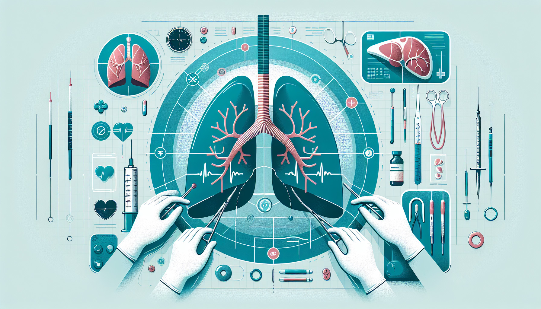
The research paper is about a study that examined the effectiveness of two types of imaging tests - immediate post-procedure computed tomography (IPP-CT) and routine one-hour chest radiography (1HR-CXR) - in spotting and managing a condition called pneumothorax in patients who have had a type of lung biopsy. Pneumothorax is when air gets into the space between the lung and the chest wall, causing the lung to collapse.
The study looked at 275 lung biopsies done on 267 patients between May 2014 and August 2021. After the biopsies, the researchers checked for pneumothorax and other complications using IPP-CT and 1HR-CXR.
The results showed that pneumothorax occurred in about 31% of the procedures. Of these, almost 90% were detected using IPP-CT and all (100%) were spotted using 1HR-CXR. A chest tube, which helps remove the air and let the lung re-expand, had to be placed in about 4% of all cases. In about 3% of the cases, pneumothorax was detected only on the 1HR-CXR and not on the IPP-CT, but none of these patients needed a chest tube.
The study also found that the likelihood of getting pneumothorax wasn’t significantly different based on the method used to close the biopsy tract, the size and type of needle used, where the needle was inserted, or the size of the lesion being biopsied. However, taking fewer biopsy samples was found to lower the risk of pneumothorax, while a longer needle tract distance increased the risk.
In conclusion, the study suggests that if pneumothorax is detected on the IPP-CT, it’s likely to persist on the 1HR-CXR and might require a chest tube. But, if no pneumothorax is spotted on the IPP-CT, the 1HR-CXR test may only be needed if the patient starts showing symptoms of pneumothorax.
FAQs
- What is the purpose of immediate post-procedure computed tomography (IPP-CT) and routine one-hour chest radiography (1HR-CXR) after a lung biopsy?
- What factors were found to be significant risk and protective factors for pneumothorax after a CT-guided percutaneous lung biopsy?
- Does the study suggest that follow-up 1HR-CXR is always necessary after a CT-guided percutaneous lung biopsy?
Doctor’s Tip
A helpful tip a doctor might tell a patient about lung biopsy is to closely follow post-procedure instructions, including monitoring for symptoms of pneumothorax such as chest pain, shortness of breath, or difficulty breathing. It is important to report any concerning symptoms to your healthcare provider immediately to ensure prompt evaluation and appropriate management.
Suitable For
Patients who are typically recommended for lung biopsy include those with suspected lung cancer, suspected infections such as tuberculosis or fungal infections, interstitial lung disease, sarcoidosis, and other lung diseases. Additionally, patients with lung nodules or masses that need to be further evaluated for diagnosis and staging may also undergo lung biopsy.
Timeline
Before lung biopsy: The patient undergoes initial consultation with a healthcare provider to discuss the need for a lung biopsy, potential risks and benefits, and to provide informed consent. Pre-procedure tests, such as blood work and imaging studies, may be performed. The patient may also be instructed on pre-procedure fasting and medication protocols.
During lung biopsy: The patient is positioned on a procedure table and a local anesthetic is administered to numb the skin. A needle or biopsy instrument is guided into the lung tissue using imaging guidance, such as CT scan. Tissue samples are collected for analysis. The patient may experience mild discomfort or pressure during the procedure.
After lung biopsy: The patient is monitored for a period of time post-procedure to check for any immediate complications, such as pneumothorax (collapsed lung) or bleeding. Immediate post-procedure CT scan may be performed to assess for pneumothorax. Routine one-hour chest radiography may also be done to further evaluate for complications. Depending on the findings, the patient may require additional monitoring, treatment (e.g. chest tube placement), or discharge instructions.
Follow-up: The patient may be advised to follow up with their healthcare provider for further evaluation of biopsy results and to monitor for any delayed complications, such as delayed pneumothorax. Any symptoms or concerns should be promptly reported to the healthcare team.
What to Ask Your Doctor
- What is the purpose of a lung biopsy and why is it being recommended for me?
- What are the risks and potential complications associated with a lung biopsy, such as pneumothorax?
- How will the procedure be performed and what can I expect during and after the biopsy?
- Will I need to have immediate post-procedure computed tomography (IPP-CT) and routine one-hour chest radiography (1HR-CXR) to monitor for pneumothorax?
- What are the signs and symptoms of pneumothorax that I should watch out for after the biopsy?
- How will pneumothorax be managed if it occurs, and when might a chest tube be necessary?
- Are there any specific factors, such as the needle diameter or access site, that could increase my risk of developing pneumothorax during the biopsy?
- How soon after the biopsy should I follow up with you to check for any complications or concerns?
- Are there any alternative procedures or imaging techniques that could be considered instead of a lung biopsy?
- What are the potential outcomes and benefits of having a lung biopsy in my specific case?
Reference
Authors: Weinand JT, du Pisanie L, Ngeve S, Commander C, Yu H. Journal: PLoS One. 2023 Apr 19;18(4):e0284145. doi: 10.1371/journal.pone.0284145. eCollection 2023. PMID: 37075048