Our Summary
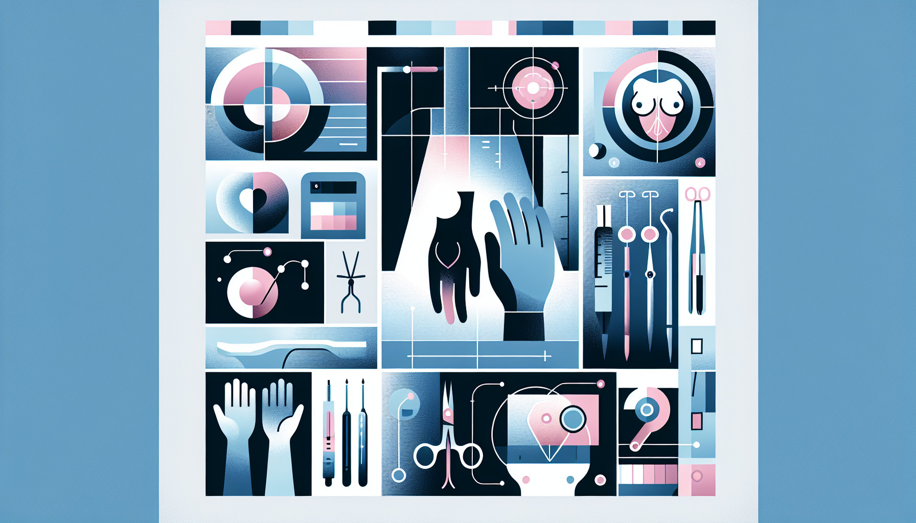
This research paper discusses the increasingly common use of digital breast tomosynthesis (DBT) - a 3D breast imaging technique - in clinical practice. Specifically, it looks at how DBT is used to guide biopsies of calcifications, which are tiny deposits of calcium in the breast that can be a sign of cancer. The authors provide a detailed guide on how to perform these biopsies, along with advice on how to handle any challenges that may come up during the process.
FAQs
- What is digital breast tomosynthesis (DBT) and how is it used in clinical practice?
- What are calcifications in the breast and how can they be a sign of cancer?
- What are some potential challenges that may come up during a breast biopsy guided by DBT?
Doctor’s Tip
One helpful tip a doctor might tell a patient about breast biopsy is to make sure to follow any pre-procedure instructions given by the healthcare provider, such as avoiding aspirin or blood thinners before the biopsy. It is also important to communicate any concerns or questions with the healthcare team before the procedure to ensure a smooth and successful biopsy process.
Suitable For
Patients who are typically recommended for breast biopsy include those with suspicious findings on mammograms, such as masses, areas of abnormal density, or calcifications. Additionally, patients with a palpable lump in the breast, changes in the skin or nipple, or abnormal results on other imaging tests may also be recommended for biopsy. It is important to note that not all patients with these findings will have cancer, but a biopsy is necessary to determine the cause of the abnormality and to guide further treatment if needed.
Timeline
Before the breast biopsy:
- The patient may undergo a screening mammogram, which may detect suspicious findings such as calcifications.
- Additional imaging tests, such as ultrasound or MRI, may be performed to further evaluate the area of concern.
- The patient may meet with a radiologist to discuss the need for a biopsy and the risks and benefits of the procedure.
- Consent for the biopsy procedure may be obtained from the patient.
- The patient may be instructed to avoid taking certain medications, such as blood thinners, before the biopsy.
During the breast biopsy:
- The patient will be positioned on a table and the skin over the biopsy site will be cleaned and numbed with a local anesthetic.
- A small incision may be made in the skin, and a biopsy needle will be inserted to collect tissue samples.
- The tissue samples will be sent to a pathology lab for analysis.
- The entire procedure typically takes about 30-60 minutes.
After the breast biopsy:
- The patient may experience some mild discomfort or bruising at the biopsy site.
- Results of the biopsy are typically available within a few days to a week.
- The patient will meet with their healthcare provider to discuss the results and determine any further treatment or follow-up care that may be needed.
- If cancer is detected, the patient may be referred to a breast surgeon or oncologist for further evaluation and treatment.
- If the biopsy results are benign, the patient may be advised to continue with regular breast cancer screening.
What to Ask Your Doctor
- What type of breast biopsy is recommended for my specific situation?
- What are the risks and benefits of having a breast biopsy?
- How will the biopsy be performed and what can I expect during the procedure?
- Will I need to undergo any additional imaging tests before or after the biopsy?
- How long will it take to receive the results of the biopsy?
- What are the potential complications of the biopsy procedure?
- What follow-up care or monitoring will be necessary after the biopsy?
- Are there any alternative options to consider besides a biopsy?
- How experienced is the healthcare provider who will be performing the biopsy?
- Are there any specific instructions or precautions I need to follow before and after the biopsy procedure?
Reference
Authors: Choudhery S, Anderson T, Valencia E. Journal: Clin Imaging. 2021 Apr;72:83-90. doi: 10.1016/j.clinimag.2020.11.035. Epub 2020 Nov 14. PMID: 33217675