Our Summary
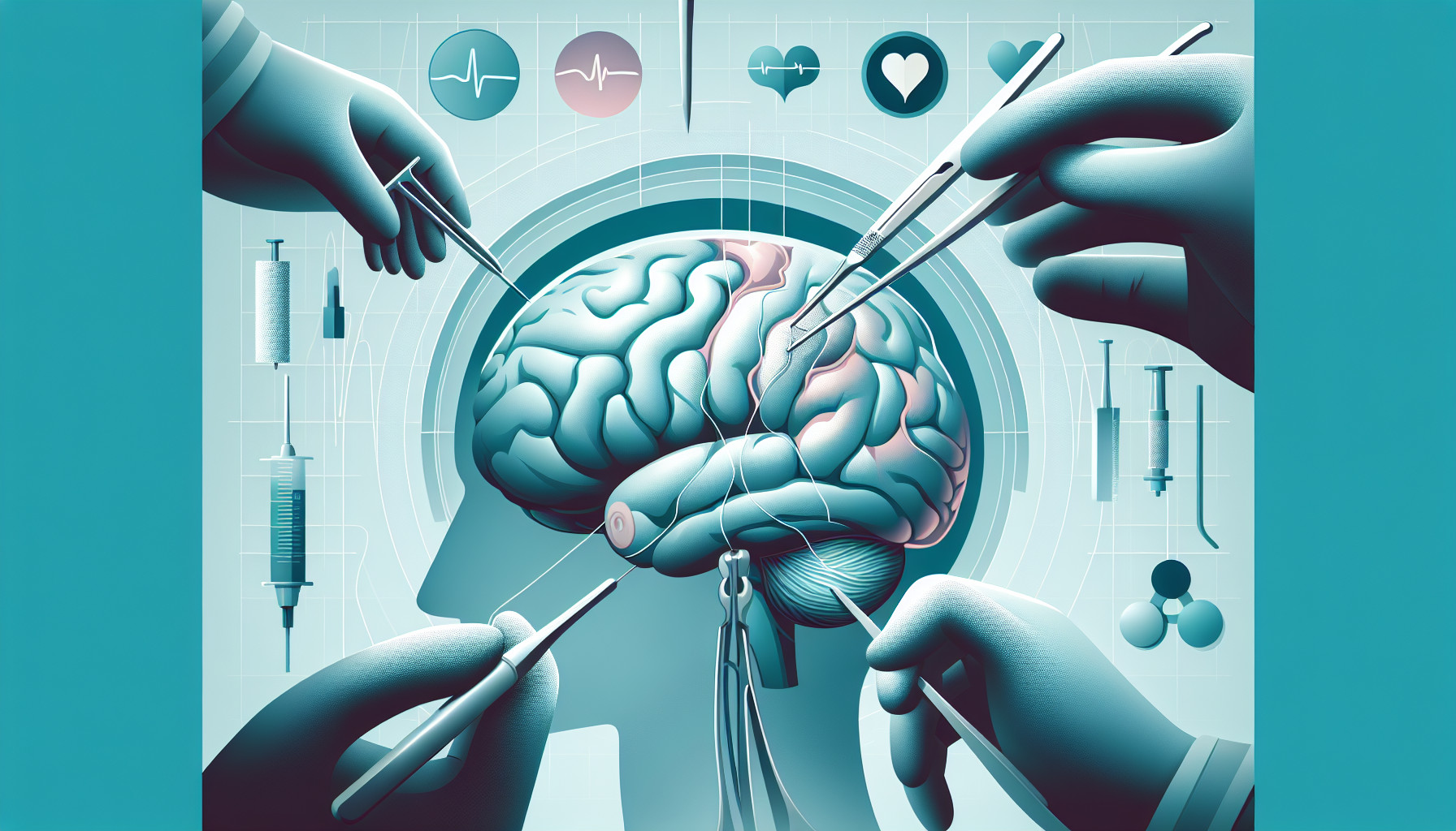
This research paper discusses a study examining the size of surgical openings, or craniotomies, needed for the placement of subdural grid electrodes in patients with epilepsy. Traditionally, large openings were made, but recently, surgeons have been moving towards smaller, personalized operations.
In a three-year period, 58 epilepsy patients had these smaller, custom-made craniotomies for electrode placement. The researchers then measured the size of these openings, the number of electrodes used, and whether the removal of the epileptic focus was successful. They compared these measurements to those from 16 patients who had the traditional, larger craniotomies.
They found that the smaller, customized craniotomies were just as successful for electrode placement. On average, these openings were 28 square centimeters, and they allowed for the placement of an average of 55 electrodes. For those with epilepsy in the temporal lobe, even smaller openings were used. Compared to the traditional method, these openings were significantly smaller, the operation time was shorter, and there were fewer complications.
Their findings suggest that smaller, personalized craniotomies are just as effective for the placement of subdural grid electrodes in epilepsy patients. They advocate for the wider use of these minimally invasive procedures in epilepsy surgery.
FAQs
- What is the average size of the smaller, custom-made craniotomies in the study?
- How do the results of the smaller, personalized craniotomies compare to the traditional, larger ones in terms of operation time and complications?
- Why do the researchers advocate for the wider use of smaller, personalized craniotomies in epilepsy surgery?
Doctor’s Tip
A doctor might tell a patient undergoing a craniotomy for epilepsy surgery that smaller, personalized openings for electrode placement can be just as effective as larger traditional openings. These smaller openings can lead to shorter operation times and fewer complications. It is important to discuss with your doctor the best approach for your specific case.
Suitable For
Patients who are typically recommended for a craniotomy include those with epilepsy who require the placement of subdural grid electrodes for monitoring and potentially treating their condition. The study discussed in this research paper specifically focused on epilepsy patients who underwent craniotomies for electrode placement, but craniotomies may also be recommended for other conditions such as brain tumors, aneurysms, traumatic brain injuries, and other neurological disorders.
In the case of epilepsy patients, craniotomies may be recommended when other treatment options have failed to adequately control seizures. The placement of subdural grid electrodes allows for the precise mapping of the epileptic focus in the brain, which can help guide surgical intervention to remove or treat the affected area. Patients who undergo craniotomies for epilepsy surgery may have tried multiple medications and other therapies without success, and their seizures may be significantly impacting their quality of life.
Overall, patients who are recommended for a craniotomy are those who can potentially benefit from surgical intervention to address their underlying neurological condition. The decision to undergo a craniotomy is typically made after careful evaluation by a multidisciplinary team of healthcare providers, including neurologists, neurosurgeons, and other specialists, to determine the most appropriate course of treatment for the individual patient.
Timeline
Before a craniotomy, a patient may experience symptoms of their underlying condition, such as seizures in the case of epilepsy. They may undergo various diagnostic tests, consultations with specialists, and discussions with their healthcare team to determine if surgery is the best option for them. The patient will also undergo pre-operative preparations, such as blood tests, imaging studies, and medication adjustments.
During the craniotomy procedure, the patient will be put under general anesthesia, and the surgeon will make an incision in the scalp and create a small opening in the skull to access the brain. In the case of epilepsy surgery, subdural grid electrodes may be placed on the surface of the brain to monitor and map the areas responsible for seizures. The electrodes are then connected to a monitoring system to record brain activity.
After the craniotomy, the patient will typically stay in the hospital for a few days for monitoring and recovery. They may experience pain, swelling, and discomfort at the surgical site, as well as potential side effects from anesthesia. The healthcare team will closely monitor the patient for any complications and provide post-operative care instructions.
In the weeks and months following the craniotomy, the patient will undergo follow-up appointments with their healthcare team to assess their recovery and monitor their progress. They may also undergo additional treatments or therapies as needed. Over time, the patient should experience improvements in their symptoms and quality of life, with the goal of better managing or resolving their underlying condition.
What to Ask Your Doctor
- What is the size of the craniotomy that will be performed for my surgery?
- How many electrodes will be placed during the procedure?
- What are the potential risks and complications associated with the craniotomy?
- How long is the recovery time expected to be following the surgery?
- What is the success rate of using smaller, personalized craniotomies for electrode placement in epilepsy patients?
- How does the use of smaller craniotomies compare to traditional, larger openings in terms of outcomes and complications?
- Will I need any additional follow-up procedures or treatments after the craniotomy?
- What is the expected outcome in terms of seizure control following the surgery?
- Are there any alternative surgical approaches or techniques that could be considered for my case?
- How experienced is the surgical team in performing smaller, personalized craniotomies for epilepsy patients?
Reference
Authors: Schneider UC, Oltmanns F, Vajkoczy P, Holtkamp M, Dehnicke C. Journal: Stereotact Funct Neurosurg. 2019;97(3):160-168. doi: 10.1159/000501235. Epub 2019 Jul 30. PMID: 31362296