Our Summary
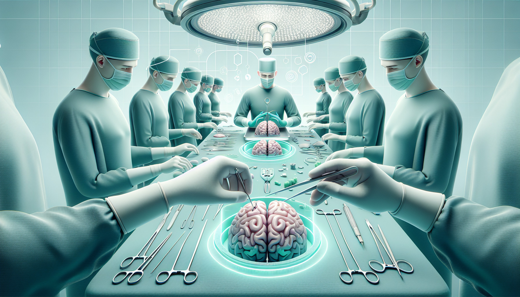
This research paper talks about a new method of performing brain surgery on small animals, which does not require the use of traditional tools like a brain atlas or stereotaxic frame. Instead, it uses a high-contrast MRI brain scan of the animal and a micro-CT scanner.
The process includes attaching tiny markers to the animal’s skull and then scanning the head using the CT scanner. The MRI and CT images are then matched using standard image processing software. The areas in the brain where recordings are to be made are marked on the MRI image.
During surgery, the animal’s head is kept stable in a suitable position, and the probe’s location and angle are tracked using a multi-camera system. The researchers have created software that uses the markers on the skull to match the CT/MRI 3D model with the surgical positioning system. The software then guides the surgeon about how to move the probe to reach the marked areas in the brain.
The researchers tested this method on owls, quails, and mice, proving its flexibility. The technique allows the creation of insertion tracks that connect two points in the brain. It is particularly beneficial for research in non-standard and novel animal models.
FAQs
- What tools are used in the new method of performing brain surgery on small animals, as opposed to traditional methods?
- How does the new method guide the surgeon during surgery?
- What benefits does the new method of brain surgery offer for research in non-standard and novel animal models?
Doctor’s Tip
In terms of brain surgery, a doctor might tell a patient to follow all pre-operative instructions carefully, such as fasting before surgery and avoiding certain medications. They may also advise the patient to discuss any concerns or questions they have with their surgical team beforehand. Additionally, the doctor may remind the patient to follow all post-operative care instructions, such as taking prescribed medications, attending follow-up appointments, and avoiding certain activities that could interfere with the healing process. It is important for the patient to communicate openly with their medical team throughout the entire process.
Suitable For
Typically, patients who are recommended for brain surgery are those with conditions such as brain tumors, epilepsy, traumatic brain injury, cerebral aneurysm, arteriovenous malformation, Parkinson’s disease, and other neurological disorders that cannot be treated effectively with medication or other non-invasive treatments. These patients may experience symptoms such as severe headaches, seizures, cognitive impairment, movement disorders, and other neurological deficits that significantly impact their quality of life.
In some cases, patients may also be recommended for brain surgery to implant deep brain stimulation devices for the treatment of conditions like Parkinson’s disease, essential tremor, and dystonia. Additionally, patients with certain types of psychiatric disorders, such as severe obsessive-compulsive disorder or treatment-resistant depression, may also be considered for neurosurgical interventions like deep brain stimulation or ablative procedures.
Overall, brain surgery is typically recommended for patients who have exhausted other treatment options and whose conditions are significantly impacting their daily functioning and quality of life. The decision to undergo brain surgery is made on a case-by-case basis by a multidisciplinary team of neurosurgeons, neurologists, neuroradiologists, and other specialists, taking into consideration the specific diagnosis, symptoms, overall health, and individual preferences of the patient.
Timeline
Before brain surgery:
- Patient undergoes various diagnostic tests such as MRI, CT scans, and EEG to identify the location and extent of the brain abnormality
- Consultation with neurosurgeon to discuss the surgical procedure, risks, and benefits
- Pre-operative preparation including fasting, medication adjustments, and possible shaving of the head
- Anesthesia induction and positioning on the operating table
- Placement of monitoring devices to track vital signs during surgery
During brain surgery:
- Neurosurgeon makes an incision in the scalp and creates a small opening in the skull to access the brain
- Removal or treatment of the abnormal tissue using specialized surgical tools and techniques
- Intraoperative monitoring to ensure the safety of surrounding brain structures
- Closure of the incision with sutures or staples
- Transfer to the recovery room for monitoring and observation
After brain surgery:
- Recovery in the hospital with close monitoring of vital signs and neurological status
- Pain management and medication to prevent infection
- Physical therapy and rehabilitation to regain lost function or improve outcomes
- Follow-up appointments with the neurosurgeon for post-operative care and monitoring of healing progress
- Long-term management of any residual symptoms or complications from the surgery
What to Ask Your Doctor
Some questions that a patient should ask their doctor about brain surgery include:
- What specific type of brain surgery do I need, and why is it necessary?
- What are the potential risks and complications associated with the surgery?
- What is the success rate of this type of surgery for my condition?
- What is the recovery process like, and how long can I expect to be in the hospital?
- Will I need any additional treatments or therapies after the surgery?
- What type of anesthesia will be used during the surgery?
- How experienced is the surgical team in performing this type of surgery?
- What are the alternatives to surgery, and why is surgery recommended in my case?
- How will my quality of life be affected after the surgery?
- Are there any long-term effects or limitations I should be aware of post-surgery?
It is important for patients to have a thorough understanding of the procedure, potential outcomes, and post-operative care before undergoing brain surgery.
Reference
Authors: Ron S, Beeri H, Shinover O, Tur NM, Brokman J, Engelhard B, Gutfreund Y. Journal: J Neurosci Methods. 2024 Nov;411:110272. doi: 10.1016/j.jneumeth.2024.110272. Epub 2024 Aug 28. PMID: 39209161