Our Summary
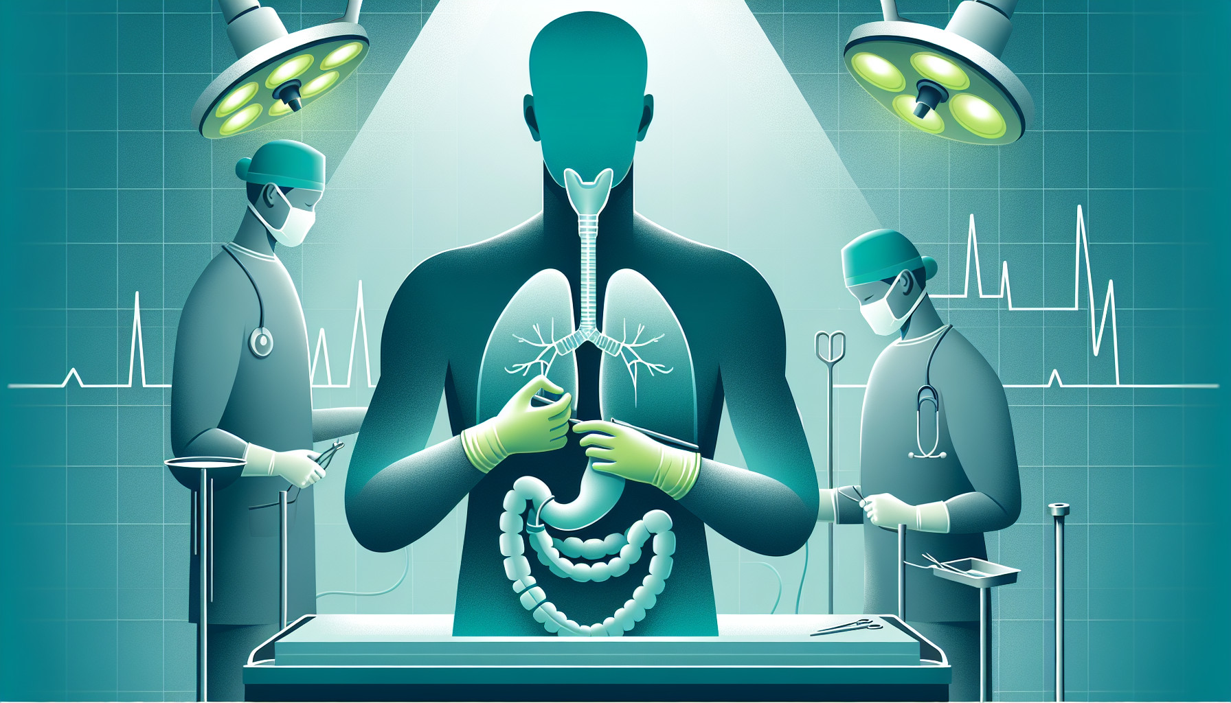
In simpler terms, this research paper discusses a surgical technique for removing cancerous cells in the esophagus (the tube that connects the throat to the stomach). However, this method carries a risk of a condition called chylothorax, which is when lymphatic fluid accumulates in the space around the lungs. This condition occurs in 5% to 20% of these types of surgeries.
To help reduce this risk, the researchers developed a new technique that uses a robot to assist the surgery. During the operation, they use a special dye (indocyanine green) that is injected into the patient’s right groin. This dye helps the surgeons better see and identify a specific duct in the chest (the thoracic duct) that is important for the removal of the lymph nodes.
This new approach can be easily applied during any similar procedure and is particularly useful when the patient is lying on their side and the surgeons need a clearer view of the thoracic duct’s structure.
FAQs
- What is the purpose of en bloc resection of the thoracic duct compartment during esophagectomy?
- What are the risks associated with this procedure and what is the incidence rate of postoperative chylothorax?
- What is the technique described in this report for intraoperative identification of the thoracic duct during robot-assisted esophagectomy?
Doctor’s Tip
A helpful tip a doctor might tell a patient about esophagectomy is to be aware of the potential risk of postoperative chylothorax, which is a rare but serious complication. Patients should follow their doctor’s recommendations for postoperative care and be vigilant for any signs of chylothorax, such as chest pain, difficulty breathing, or coughing up milky fluid. If any concerning symptoms occur, it is important to contact their healthcare provider immediately for evaluation and treatment.
Suitable For
Patients who may be recommended for esophagectomy include those with esophageal cancer, Barrett’s esophagus with high-grade dysplasia, benign esophageal strictures that are not responding to other treatments, and esophageal motility disorders such as achalasia. Additionally, patients who have failed previous treatments such as chemotherapy or radiation therapy may also be candidates for esophagectomy.
Timeline
Before esophagectomy:
- Patient undergoes various diagnostic tests such as endoscopy, biopsy, CT scans, and PET scans to confirm the presence of malignant esophageal disease.
- Patient meets with a multidisciplinary team of healthcare providers to discuss treatment options and risks, including the possibility of postoperative complications.
- Patient undergoes preoperative preparations such as fasting, bowel preparation, and medication adjustments.
- Patient may receive preoperative counseling and education on the procedure, recovery process, and potential complications.
After esophagectomy:
- Patient is closely monitored in the recovery room for immediate postoperative complications such as bleeding, infection, or respiratory issues.
- Patient may be transferred to the intensive care unit for closer observation and management of pain, nutrition, and fluid balance.
- Patient gradually resumes oral intake and starts physical therapy to regain strength and mobility.
- Patient may experience complications such as chylothorax, anastomotic leakage, or stricture formation, which require additional treatment and monitoring.
- Patient undergoes regular follow-up appointments with their healthcare team to monitor recovery progress, manage any complications, and provide ongoing support and guidance.
What to Ask Your Doctor
- What is the purpose of en bloc resection of the thoracic duct compartment during esophagectomy for malignant esophageal diseases?
- What are the potential benefits of enhancing lymph node removal in this procedure?
- What is the risk of developing postoperative chylothorax after this type of esophagectomy?
- How common is postoperative chylothorax following this procedure?
- How is the thoracic duct typically identified and managed during esophagectomy?
- Can you explain the technique of lymphangiography-guided injection of indocyanine green in the right groin for facilitating intraoperative identification and ligation of the thoracic duct?
- Are there any specific precautions or considerations that need to be taken before or during this procedure to reduce the risk of postoperative complications?
- What is the success rate of using this technique in identifying and managing the thoracic duct during esophagectomy?
- What is the recovery process like for patients who undergo this type of esophagectomy, particularly in relation to the risk of chylothorax?
- Are there any long-term implications or considerations to be aware of after undergoing en bloc resection of the thoracic duct compartment during esophagectomy?
Reference
Authors: Jardinet T, Niekel MC, Ruppert M, Hubens G, Valk JW, van Schil PE, de Maat MF. Journal: Ann Thorac Surg. 2022 Jun;113(6):e465-e467. doi: 10.1016/j.athoracsur.2021.08.034. Epub 2021 Sep 22. PMID: 34560041