Our Summary
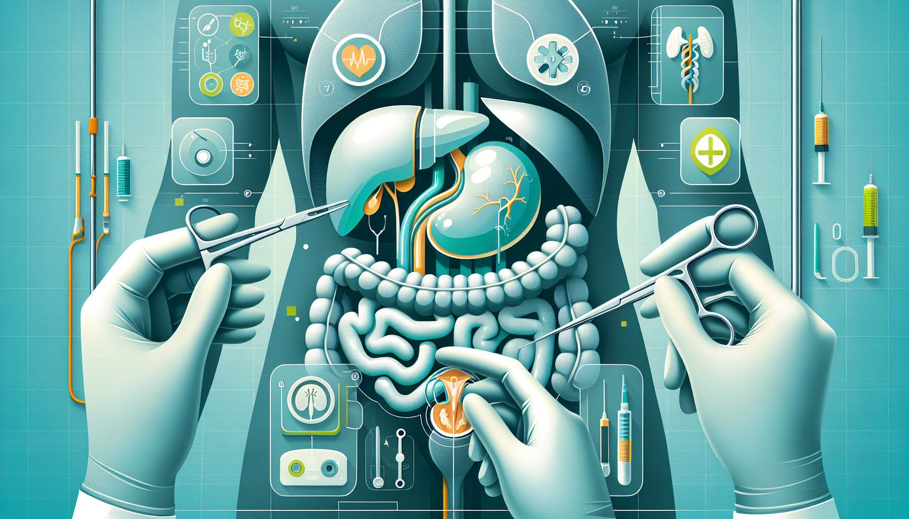
This research study explored the use of a technology called indocyanine green (ICG) fluorescence imaging during a type of surgical procedure called laparoscopic partial adrenalectomy. This surgery involves the partial removal of the adrenal glands, which are small glands located on top of each kidney. The researchers were particularly interested in how this technology could be used for patients with pheochromocytoma (a rare tumor that starts in the adrenal glands) and Cushing’s syndrome (a condition that happens when your body has too much cortisol).
ICG fluorescence imaging uses a special dye that gives off light when it is exposed to near-infrared rays, allowing doctors to see the blood flow in the adrenal glands and surrounding tissues more clearly. This is important because it helps surgeons to precisely locate the tumors and to understand how well the remaining adrenal tissue is being supplied with blood after part of the gland is removed.
The researchers tested this technology on three patients undergoing laparoscopic partial adrenalectomy for pheochromocytoma and Cushing’s syndrome. They found that ICG imaging was useful for identifying the edges of the adrenal glands and the adrenal vein, especially on the left side. It also helped them to find and remove the entire tumor. On the right side, where the anatomy is more apparent, ICG imaging did not make much difference to the operation.
The researchers concluded that ICG fluorescence imaging could be a helpful tool during partial adrenalectomy, both for guiding the surgery and for assessing the blood supply to the remaining adrenal tissue.
FAQs
- What is indocyanine green (ICG) fluorescence imaging and how is it used in laparoscopic partial adrenalectomy?
- Why is the use of ICG fluorescence imaging important during laparoscopic partial adrenalectomy for pheochromocytoma and Cushing’s syndrome?
- How effective was the use of ICG fluorescence imaging in the research study and what conclusions were drawn by the researchers?
Doctor’s Tip
A doctor might tell a patient undergoing adrenalectomy to ask about the possibility of using ICG fluorescence imaging during the surgery. This technology can help the surgeon accurately locate the tumor and ensure that the remaining adrenal tissue is well supplied with blood. It may improve the overall outcome of the procedure and reduce the risk of complications.
Suitable For
In general, patients who may be recommended for adrenalectomy include those with:
- Adrenal tumors, including adrenal adenomas, adrenal carcinomas, pheochromocytomas, and aldosterone-producing adenomas
- Cushing’s syndrome, which is caused by the overproduction of cortisol by the adrenal glands
- Conn’s syndrome, which is caused by the overproduction of aldosterone by the adrenal glands
- Adrenal metastases, which are secondary tumors that have spread to the adrenal glands from other parts of the body
It is important to note that the decision to undergo adrenalectomy is made on a case-by-case basis, taking into consideration the patient’s specific condition, overall health, and treatment goals. Consulting with an endocrinologist or a surgeon specializing in adrenal surgery is recommended to determine the best course of action for each individual patient.
Timeline
Before adrenalectomy:
- Patient undergoes diagnostic tests (e.g. blood tests, imaging studies) to confirm the presence of a tumor in the adrenal glands.
- Patient meets with an endocrinologist and surgeon to discuss treatment options and surgical risks.
- Patient may undergo medical treatment to stabilize hormone levels before surgery.
- Patient undergoes preoperative evaluations and preparations for surgery.
After adrenalectomy:
- Patient is monitored in the hospital for complications such as bleeding, infection, or hormone imbalances.
- Patient may need to take hormone replacement therapy to manage symptoms of adrenal insufficiency.
- Patient undergoes follow-up appointments with their healthcare team to monitor recovery and hormone levels.
- Patient may need to make dietary and lifestyle changes to manage any long-term effects of the surgery.
What to Ask Your Doctor
- How does ICG fluorescence imaging work and how will it benefit me during my adrenalectomy surgery?
- Are there any risks or side effects associated with using ICG dye during the procedure?
- Will ICG imaging help ensure that the entire tumor is removed during the surgery?
- How will ICG imaging help in preserving the function of the remaining adrenal tissue?
- What are the potential long-term benefits of using ICG fluorescence imaging during adrenalectomy?
- Are there any specific criteria that make a patient a good candidate for ICG imaging during adrenalectomy?
- How experienced is the surgical team with using ICG imaging during adrenalectomy procedures?
- Will ICG imaging affect the overall duration of the surgery or my recovery time?
- Are there any additional costs associated with using ICG imaging during the surgery?
- Are there any alternative imaging techniques or approaches that could be used instead of ICG fluorescence imaging during adrenalectomy?
Reference
Authors: Lerchenberger M, Gündogar U, Al Arabi N, Gallwas JKS, Stepp H, Hallfeldt KKJ, Ladurner R. Journal: Surg Endosc. 2020 May;34(5):2050-2055. doi: 10.1007/s00464-019-06985-7. Epub 2019 Jul 24. PMID: 31342258