Our Summary
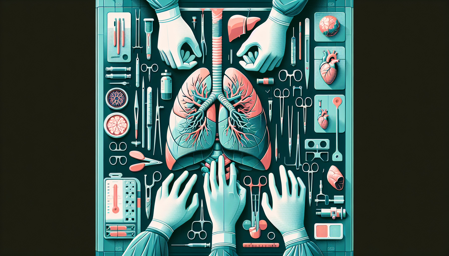
This study tested a new method called virtual-assisted lung mapping 2.0 for removing deeply located lung nodules, which are small growths in the lung that could potentially be cancerous. This technique uses a mix of dye marks and tiny coils to guide the surgeon during the operation.
This study involved 64 patients across 8 medical centers, who had nodules that were hard to see and needed to be removed. The goal was to remove the nodules with a good margin of healthy tissue around them to ensure all potential cancer was removed. The dye and coils were placed in the patients’ lungs one or two days before the surgery.
The study found that this technique was very successful, with 98.5% of nodules being successfully removed. Only 3 of the 75 coils moved out of place after being put in. No serious side effects were associated with the procedure.
This suggests that virtual-assisted lung mapping 2.0 can effectively help surgeons remove deep lung nodules, improving upon the limitations of older methods.
FAQs
- What is the new method called virtual-assisted lung mapping 2.0 used for?
- How successful was the virtual-assisted lung mapping 2.0 technique in the study?
- Were there any side effects associated with the virtual-assisted lung mapping 2.0 procedure?
Doctor’s Tip
One helpful tip a doctor might give to a patient undergoing lung resection is to ask about the possibility of using virtual-assisted lung mapping 2.0 to guide the surgery. This technique can help ensure that the nodules are removed with a good margin of healthy tissue, reducing the risk of leaving any cancerous cells behind. Be sure to discuss this option with your healthcare team to see if it is appropriate for your specific case.
Suitable For
Patients who are typically recommended for lung resection include those with lung nodules that are suspected to be cancerous, those with early-stage lung cancer, those with lung infections or abscesses, and those with lung damage from conditions such as emphysema or cystic fibrosis. This new technique of virtual-assisted lung mapping 2.0 can be particularly beneficial for patients with deeply located lung nodules that are difficult to see and remove using traditional methods.
Timeline
Before the lung resection:
- Patient undergoes imaging tests (such as CT scans) to locate the lung nodule
- Patient may undergo a bronchoscopy or biopsy to determine if the nodule is cancerous
- If the nodule needs to be removed, the patient may undergo virtual-assisted lung mapping 2.0 procedure to mark the nodule’s location
- The dye and coils are placed in the patient’s lungs one or two days before the surgery
After the lung resection:
- Surgeon uses the virtual-assisted lung mapping 2.0 technique to guide the removal of the lung nodule
- The nodule is removed along with a margin of healthy tissue to ensure all potential cancer is removed
- The success of the procedure is evaluated and any complications or side effects are monitored
- Patient undergoes recovery and follow-up care to ensure proper healing and monitor for any recurrence of the nodule or cancer
What to Ask Your Doctor
- What is the success rate of virtual-assisted lung mapping 2.0 for removing deeply located lung nodules?
- What are the potential risks or side effects associated with this technique?
- How does virtual-assisted lung mapping 2.0 differ from traditional methods of removing lung nodules?
- What is the recovery time and expected outcome after undergoing lung resection with this technique?
- Are there any specific criteria or factors that make a patient a good candidate for virtual-assisted lung mapping 2.0?
- How long after the dye and coils are placed in the lungs does the surgery typically take place?
- Will I need any additional follow-up procedures or tests after the lung resection with virtual-assisted lung mapping 2.0?
- Are there any long-term effects or considerations to be aware of after undergoing this procedure?
- How does virtual-assisted lung mapping 2.0 compare to other minimally invasive techniques for removing lung nodules?
- Are there any ongoing research or advancements in this area that I should be aware of?
Reference
Authors: Sato M, Kobayashi M, Sakamoto J, Fukai R, Takizawa H, Shinohara S, Kojima F, Sakurada A, Nakajima J. Journal: J Thorac Cardiovasc Surg. 2022 Jul;164(1):243-251.e5. doi: 10.1016/j.jtcvs.2021.09.016. Epub 2021 Sep 17. PMID: 34654560